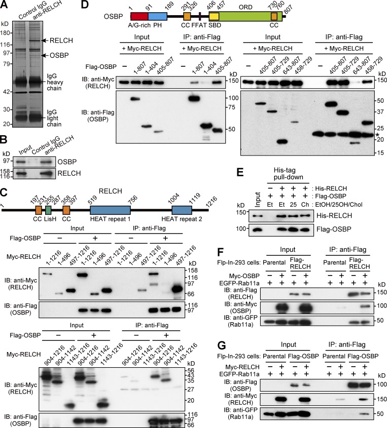
Figure 2.
OSBP is a RELCH-binding protein. (A and B) Protein G Sepharose beads bound to control IgG or the RELCH antibody were incubated with a mouse brain lysate. The precipitated samples were analyzed by SDS-PAGE and silver staining and further analyzed by mass spectrometry (A). The mass spectrometry analysis identified RELCH and OSBP, which are indicated by the arrows. The precipitated samples were analyzed by immunoblotting (IB) using the OSBP and RELCH antibodies (B). (C and D) The lysates of HEK293FT cells coexpressing a series of Flag-tagged OSBP and Myc-tagged RELCH fragments were immunoprecipitated (IP) using a Flag antibody. The samples were immunoblotted with the Myc and Flag antibodies. SBD, sterol-binding domain. In total, 0.5% and 1.5% of the input samples were loaded in upper and lower panels, respectively (C). The asterisk indicates the nonspecific bands (D). (E) A His-tag pulldown assay was performed using His-RELCH and Flag-OSBP in the presence of solvent (ethanol, EtOH), 25-OH, or cholesterol (Chol). The samples were immunoblotted with the His and Flag antibodies. (F and G) The Flag-, EGFP-, and Myc-tagged proteins were coexpressed in the Flp-In–293 cells. The lysates were immunoprecipitated using a Flag antibody, and the samples were immunoblotted with the EGFP, Myc, and Flag antibodies.

