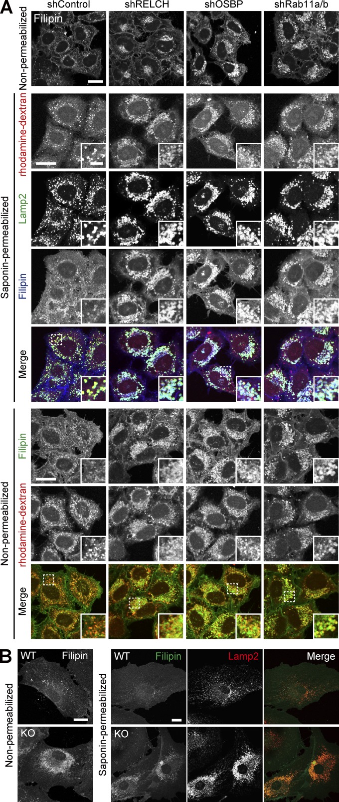
Figure 5.
Cholesterol accumulation in LEs/lysosomes after RELCH, OSBP, or Rab11 depletion. (A) The lysosomes were labeled with rhodamine-dextran in the shRNA-treated cells. The cells were stained with Filipin under the nonpermeabilized condition and Filipin and Lamp2 under the permeabilized condition. (B) Mouse tail–tip fibroblasts derived from WT and RELCH KO mice were stained with the Lamp2 antibody and/or Filipin. Bars: (main images) 20 µm; (insets) 2 µm.

