Figure 7.
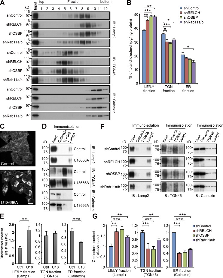
RELCH, OSBP, and Rab11 depletion results in less cholesterol accumulation in the TGN and ER. (A and B) The homogenates from the shRNA-expressing HeLa cells were fractionated using a Histodenz step density gradient. (A) The fractions were immunoblotted (IB) with antibodies against TGN46, calnexin, and Lamp2. (B) Percentages of cholesterol (µg/mg protein) in the TGN (fractions 4–6), LEs/lysosomes (LE/LY; 7–9), or ER (10 and 11) in the total fractions are shown in the bar graph. (C) HeLa cells were treated with 2 µg/ml U18666A for 16 h and stained with Filipin. Bar, 20 µm. (D–G) Immunoisolation of ER, TGN, and LE/lysosome from the PNS derived from the U18666A-treated (D and E) or RELCH-, Rab11a/b-, and OSBP-depleted cells by shRNAs (F and G). ER, TGN, and LE/lysosome membranes were isolated using calnexin, TGN46, and Lamp1 antibodies, respectively. (D and F) The isolated samples were immunoblotted with calnexin, TGN46, and Lamp2 antibodies. (E and G) Quantification of the cholesterol content in the isolated membranes (relative to the control samples). Data are expressed as means ± SEM from at least three independent experiments. *, P < 0.05; **, P < 0.01; ***, P < 0.001 (relative to the control; two tailed Student’s t tests). Representative data from three independent experiments are shown in A, D, and F.

