Figure 1.
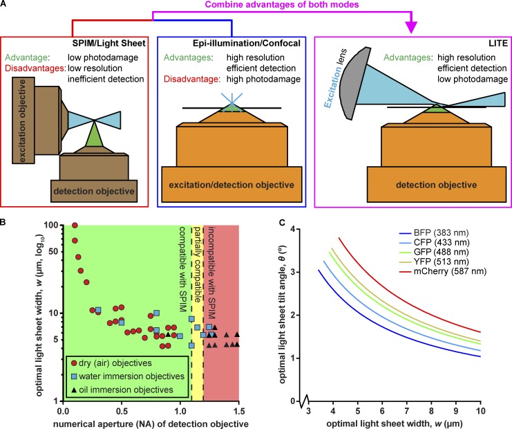
Rationale and theory behind LITE. (A) We combined low photodamage of SPIM/LSFM (left) with high-NA objectives (orange) of epi-illumination/confocal microscopy (center) to create LITE (right). LITE tilts a cylindrical lens (gray) to focus a laser (blue) into a sheet onto the coverslip surface (black line). (B) Scatterplot of calculated optimal light-sheet width for LITE, based on Eq. 7, for 90 commercially available objectives (plotted by increasing NA). Green area: objectives that can/have been used with existing live-cell SPIM/LSFM technologies. Yellow area: objectives that have been used with live-cell SPIM/LSFM by means of unconventional geometries. Red area: objectives previously incompatible with live-cell SPIM/LSFM. (C) Theoretical optimal light sheet width, w, as a function of optimal sheet tilt angle, θ. Ideal light sheet parameters are traced for five common fluorescent proteins: blue fluorescent protein (BFP), cyan fluorescent protein (CFP), green fluorescent protein (GFP), yellow fluorescent protein (YFP), and monomeric Cherry (mCherry). Wavelengths of excitation light plotted in C correspond to maximal-absorption wavelength of each protein.

