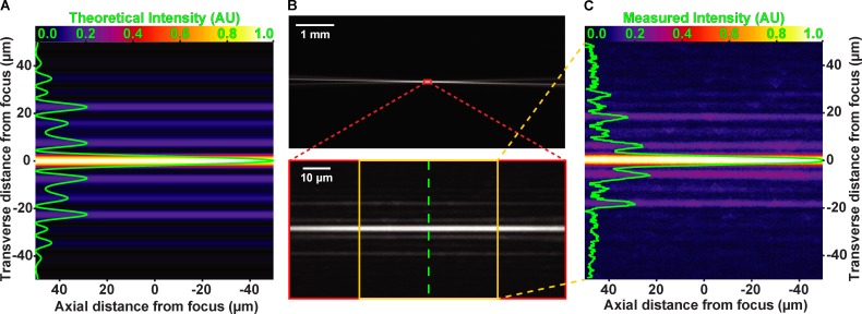
Figure 2.
Experimental verification of theoretical light sheet formation. All images in show light sheet from the side. (A) Theoretical interference pattern at cylindrical lens focus. Image has been false-colored by “Fire” lookup table in Fiji (scale at top). Transverse (across sheet width) intensity line scan is overlaid in green. FWHM of the central peak is predicted to measure 4.3 µm. (B) Low-magnification image (upper) of light sheet focusing into fluorescent media. High-magnification image (lower) of cylindrical lens focal region. Green line indicates location of transverse line scan of measured intensity. (C) Subset of the image in lower portion of B (between the yellow lines). Line scan (green line), scale, and coloration are consistent with predicted pattern in A. Measured FWHM of the central sheet is 4.3 µm, in agreement with the theoretical prediction.

