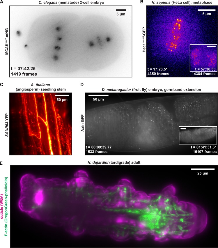
Figure 5.
Representative LITE fluorescent images taken of a variety of model organisms. The organisms include C. elegans (A), H. sapiens (B), A. thaliana (C), D. melanogaster (D), and H. dujardini (E). Fluorescent constructs imaged in each organism are delineated to left of each representative image. Images presented in A, B, and D are taken from the full movies available in Videos 3, 6, and 4, respectively. Images in C and E are static images taken from 3D z-stacks, which are presented fully in Videos 2 and 5, respectively. Insets in B and D show images taken from later time points (identically scaled) to show low photobleaching. All 2D images presented are maximal-intensity projections of a z-series.

