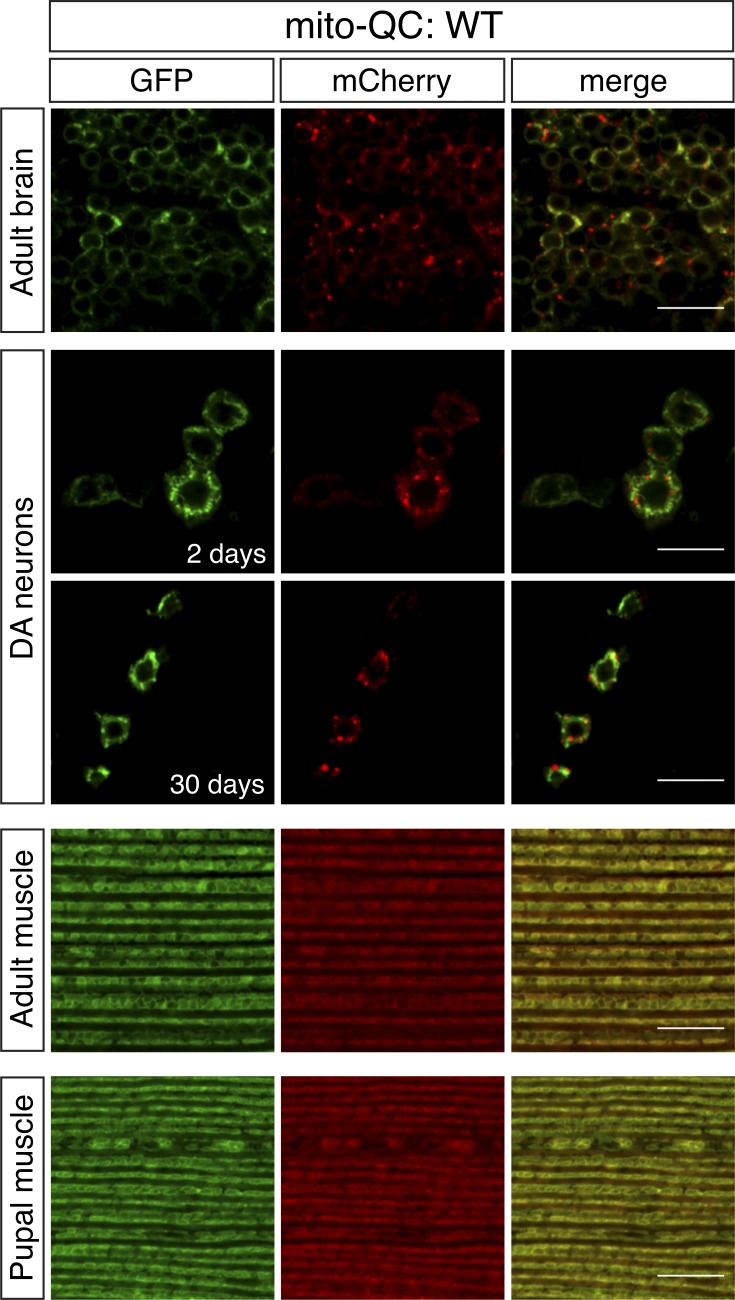
Figure 3.
Mitolysosome analysis with mito-QC in adult tissues. Confocal microscopy analysis of mito-QC reporter in WT adult brain (2 d old), DA neurons at 2 and 30 d old, flight muscle (2 d old), and preadult (pupal) flight muscle, as indicated. Mitolysosomes are evident as GFP-negative/mCherry-positive (red-only) puncta. Quantification of mitolysosomes is shown in Figs. 4 and 5. Genotypes analyzed were UAS-mito-QC/+;nSyb-GAL4/+ (adult brain), UAS-mito-QC/+;TH-GAL4/+ (DA neurons), and Mef2-GAL4/UAS-mito-QC (adult and pupal muscle). Bars, 10 µm.

