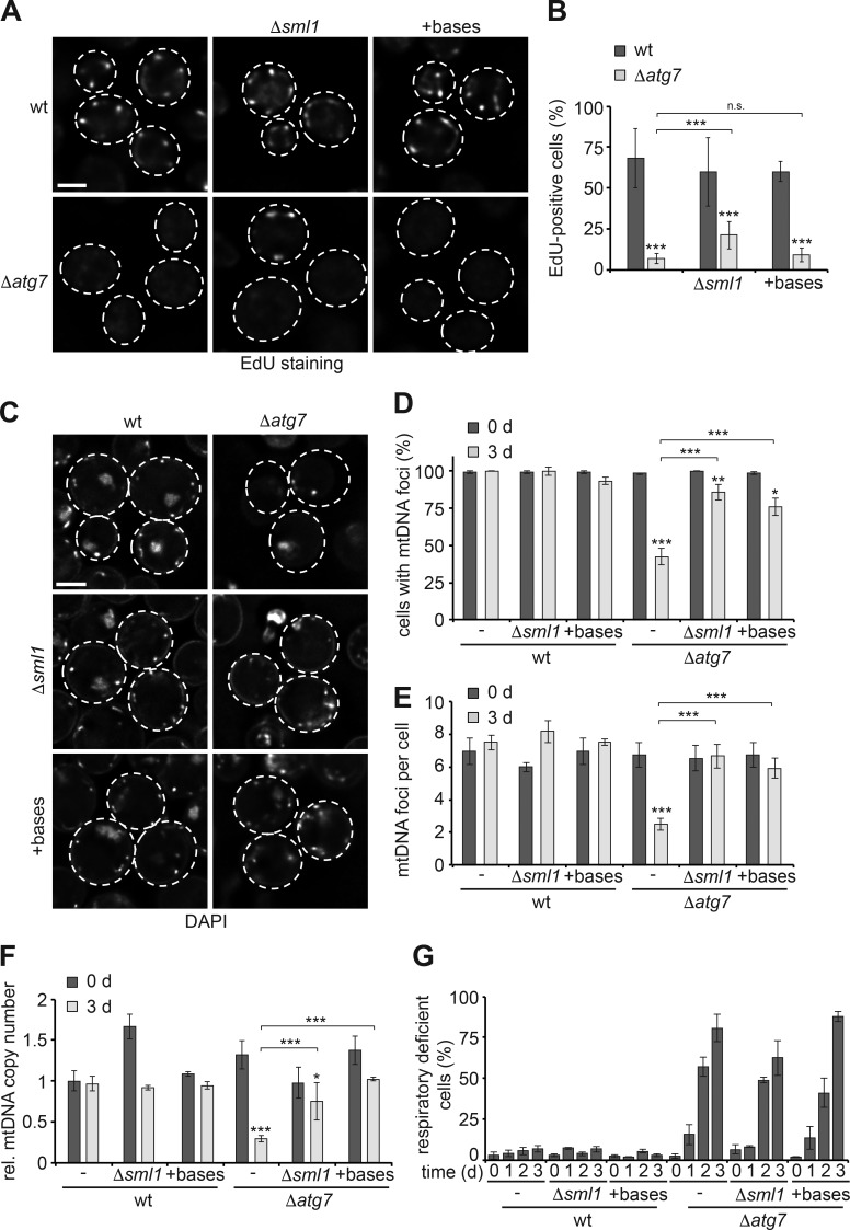
Figure 2.
Nucleotide availability limits mtDNA stability in autophagy-deficient cells during starvation. (A and B) mtDNA synthesis in dependence of nucleotide levels. WT, Δsml1, Δatg7, and Δsml1Δatg7 cells or WT and Δatg7 cells supplemented with nucleobases were assessed after 1 d of starvation as described in Fig. 1 (A and B). Data are means ± SD (n ≥ 3; 150 cells). (C–E) Increased nucleotide levels stabilize mtDNA in the absence of autophagy. WT, Δsml1, Δatg7, and Δsml1Δatg7 cells or WT and Δatg7 cells supplemented with nucleobases were analyzed by DAPI staining and fluorescence imaging as described in Fig. 1 (C–E). Data are means ± SD (n = 3; ≥75 cells). (F) Increased nucleotide levels stabilize mtDNA in autophagy-deficient cells during starvation. Cells were treated as described in C and analyzed by quantitative PCR as described in Fig. 1 F. Data are means ± SD (n ≥ 3). (G) Increased nucleotide levels do not sustain functional integrity of mtDNA in autophagy mutants. Cells treated as described in C were analyzed as described in Fig. 1 G. Data are means ± SD (n = 3). Dashed lines indicate cell boundaries. Bars, 2 µm. t tests: *, P < 0.05; **, P < 0.01; ***, P < 0.001; n.s., not significant. Rel., relative.

