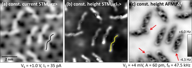
Figure 3.
Single 6P molecules imaged with STM and nc-AFM at 5 K. (a, b) Molecules adopt an asymmetric W shape, as indicated in black/yellow. (c) Constant-height nc-AFM (unknown tip) reveals an anisotropic pattern along the molecules’ long axis. One end of each 6P (red arrows) shows a clearly discerned phenyl ring.

