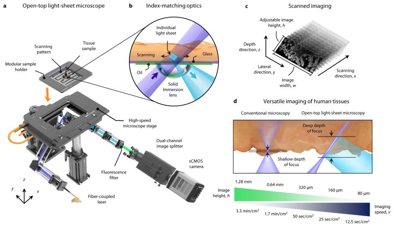
Figure 1. Open-top light-sheet microscope for clinical pathology.
a, An illumination light sheet enters the bottom surface of a tissue sample at an oblique 45-deg angle (purple). The specimen(s) is placed on a modular glass-plate sample holder, which is inserted into a two-axis translation stage and scanned in a serpentine pattern of volumetric image strips to enable 3D imaging over a large lateral extent. Fluorescence emission (cyan), which is generated along the light sheet, is collected in the orthogonal direction by an objective lens. The fluorescence signal is then transmitted through an emission filter (green) and a dual-channel image splitter (for 2 color imaging) before being imaged onto a high-speed sCMOS camera. b, To provide aberration-free imaging, a solid immersion lens (SIL) and oil layer are used for refractive-index matching of both the illumination and collection beams into and out of the glass plate and tissue sample. c, As the sample is translated in the primary scanning direction, x, oblique 2D light-sheet images with a width, w, and adjustable height, h, are captured in succession to form a 3D imaging volume. d, In contrast to conventional microscopy methods that have a shallow fixed depth of focus and slow 3D imaging rates, the deep depth of focus and adjustable vertical field of view of the open-top light-sheet microscope makes it optimal for both rapid microscopy of irregular/tilted tissue surfaces, and deep volumetric microscopy of clinical specimens. The imaging speeds shown correspond to acquiring single-channel images with height, h.

