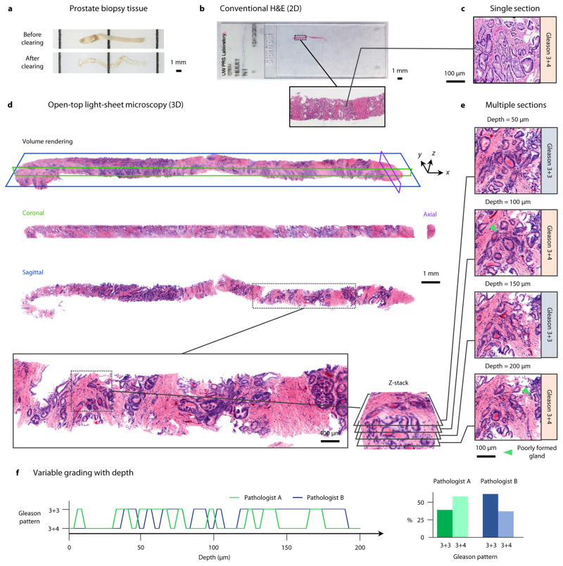
Figure 4. Volumetric dual-channel imaging of a cleared human biopsy.
a, A 2-cm long by 1-mm diameter human prostate core-needle biopsy, before and after optical clearing (overnight procedure). b, A conventional H&E-stained slide of a single 5-μm thick section of the biopsy sample. c, A high-magnification view of a Gleason 3+4 carcinoma region from the histology slide. d, The prostate biopsy was stained with DRAQ5 and eosin, and false-colored to mimic the traditional H&E color palette. A volume rendering of the biopsy is shown, which can be digitally “sectioned” into orthogonal 2D cross sections in the coronal (green), axial (purple), and sagittal (blue) planes. e, A high-magnification view of one sagittal region of interest, visualized at multiple depths (5 μm increments) to a depth of 200 μm. Gleason grading is found to differ between 3+3 and 3+4 due to tangential sectioning artifacts creating the appearance of poorly formed glands (inset arrows). f, Grading of the region of interest as a function of depth (5-μm increments) by two pathologists. When assessing individual depths, the grading varies between 3+3 (40% for Pathologist A, 62.5% for Pathologist B) and 3+4 (60% for Pathologist A, 37.5% for Pathologist B), with inter-observer agreement of 47.5%. However, after comprehensive review of the entire volumetric dataset, both pathologists agreed upon an unambiguous diagnosis of Gleason 3+3 based on the observation that structures which appeared to be “poorly formed” pattern 4 glands were tangential-sectioning artifacts of well-formed pattern 3 glands.

