. 2018 Mar 22;7:e32120. doi: 10.7554/eLife.32120

Figure 1. Propagation block by cuts in the dorsal alligator atrioventricular canal reveals a specialized atrioventricular conduction system.

(A-B) Cuts in a small region of the dorsal atrioventricular canal induces atrioventricular block (dorsal cut n = 3 and ventral cut n = 3). (C-D) Extensive cuts to the ventral and lateral atrioventricular canal do not induce atrioventricular block. (E) In vivo and ex vivo (in bath) QRS duration was not different (n = 7). (F) In bath optical mapping of ventricular activation revealed epicardial breakthrough of the impulse deep in the ventricle (n = 6). (G) The maximum rate of depolarization (red star) occurred earlier at position S than at position LV and RV of panel F (n = 6). (H-I) Optical recordings from the inside of the heart (H) show earliest activation in the septum (n = 1) (I). AVC, atrioventricular canal; LA, left atrium; LV, left ventricle; RA, right atrium; RV, right ventricle; s, ventricular sulcus; VS, ventricular septum.

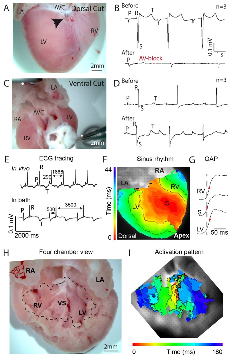
Figure 1.

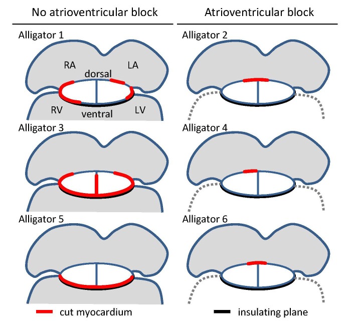
Figure 1—figure supplement 1. Overview of cuts (red) to the atrioventricular junction and the extent of collagen (black) in the six juvenile American alligators where atrioventricular conduction was assessed and where optical mapping was performed.

Figure 1—figure supplement 1.

The positions of the cuts were based on visual inspection of the fixed hearts and confirmed by histology. LA, left atrium; LV, left ventricle; RA, right atrium; RV, right ventricle.
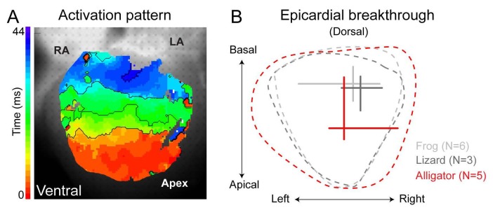
Figure 1—figure supplement 2. Ventricular activation patterns in reptiles.

Figure 1—figure supplement 2.

(A) Shows the ventricular activation pattern at the ventral side of the alligator heart (n = 6). (B) Summary of the earliest epicardial breakthrough on the dorsal ventricular surface of the American alligator compared to the green anole lizard and the Xenopus frog (lizard and frog modified from [Jensen et al., 2012]). Note that the position of breakthrough in the alligator heart is located closer to the apex compared to the frog and the lizard. RA, right atrium; LA, left atrium.