. 2018 Mar 22;7:e32120. doi: 10.7554/eLife.32120

Figure 6. Evolutionary conservation of the trabeculated and compact ventricular wall.

(A) The ventricular septum of the alligator is highly trabeculated (also Figure 6—figure supplement 1). Yet the alligator septum does not express the trabecular marker Nppb (n = 5). (B) The developing septum of alligators and mouse is empty for Nppb, in contrast to chicken (HH28 and HH42), whereas the developing septum of alligators and chicken is highly trabeculated, in contrast to the septum in mouse which is compact. The compact wall of both alligator (n = 1), caiman (n = 1), and mouse expresses Hey2. Arrowheads (white, no stain; red, stain); c, coronary artery; LV, left ventricle; RV, right ventricle.

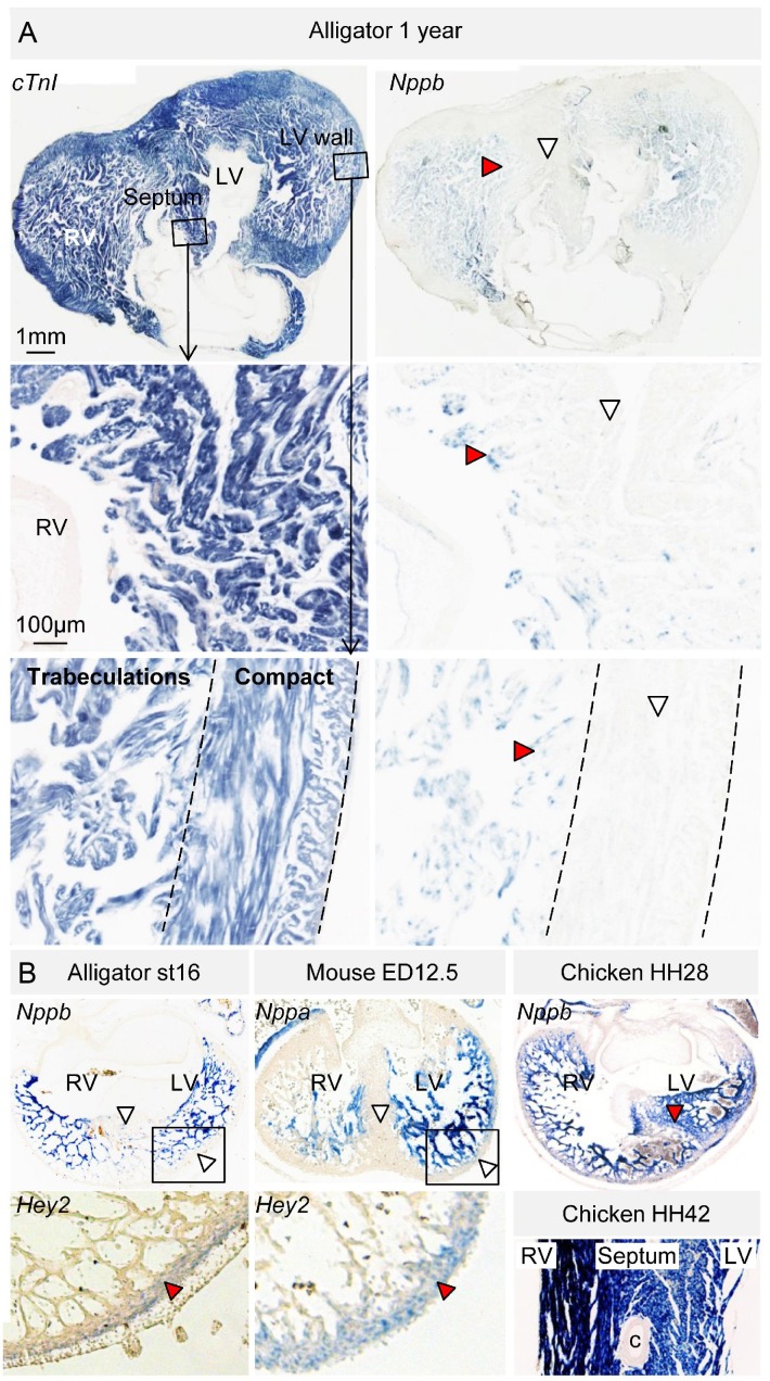
Figure 6.

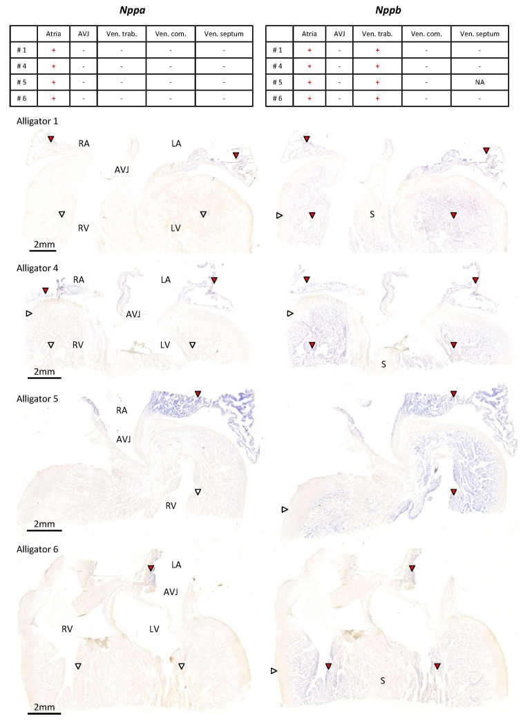
Figure 6—figure supplement 1. Expression of Nppa and Nppb in the ventricular base of 1 year old alligators.

Figure 6—figure supplement 1.

The tables summarize the pattern of expression in the images below. Nppa was only found in atrial myocardium (red arrowheads) and was not detected in the atrioventricular junction (AVJ). Nppb was expressed in the atria and the trabeculated myocardium of the ventricle (red arrowheads) but was not detected from the compact wall of the ventricle (white arrowheads) and the core of the ventricular septum (S).
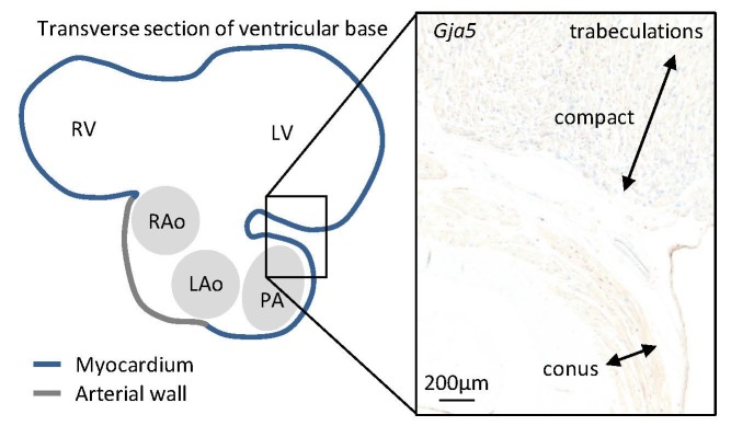
Figure 6—figure supplement 2. In the juvenile alligators, Gja5 expression was found throughout the trabecular ventricle and in the substantial compact myocardium (more than 0.5 mm thick).

Figure 6—figure supplement 2.

Only the myocardial outflow tract (the conus arteriosus) expressed very little Gja5.