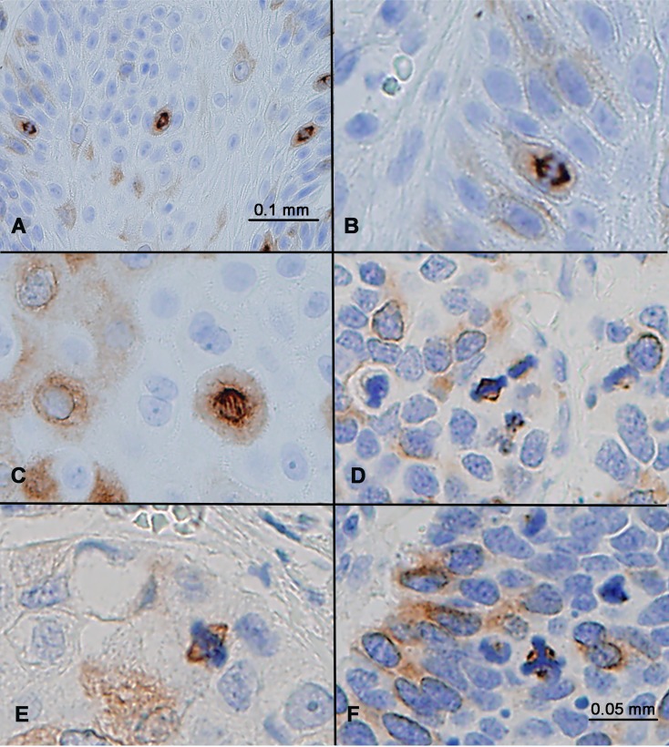
Figure 4. RHAMM expression in mitotic cells.
(A) Squamous cell carcinoma showed strong RHAMM staining of all mitotic figures, in addition to some cells with weak cytoplasmic staining. (B–F) RHAMM staining in mitotic anaphase (B, C), metaphase (D, E), and in a tumor cell with abnormal tripolar mitosis (F) in a small cell lung carcinoma.

