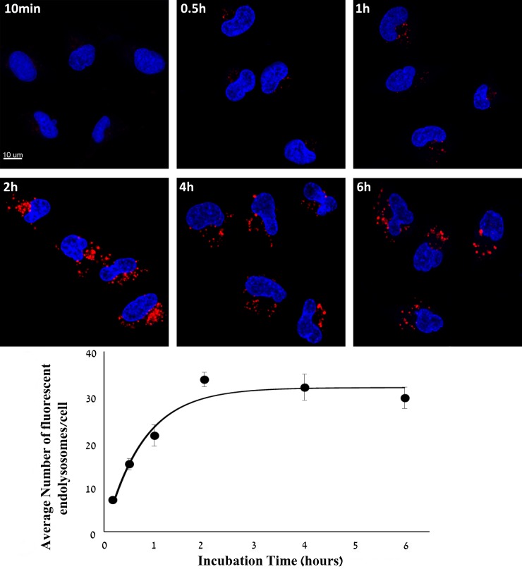
Figure 2. Kinetic study of cellular internalization of S15-APT QDs into A549 cells.
A549 cells were incubated with 50 nM S15-APT QDs for 10 min, 0.5 h, 1 h, 2 h, 4 h, and 6 h at 37° C. Nuclei were stained with 2 μg/ml Hoechst 33342. Fluorescence confocal microscopy was performed using an inverted confocal microscope (Zeiss LSM 710) at ×630 magnification. Quantification of the average number of endolysosomes per cell was determined using Imaris Software. The red fluorescence channel was defined between 10-100 for all presented images. Fitting the dependence of the average number of fluoresent endolysosomes/cell (Y(t)) to the incubation time (t) was performed by nonlinear curve fitting (Eq. 1).

