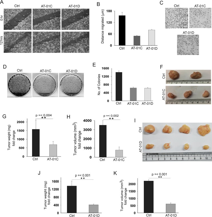
Figure 7. AT-01C and AT-01D exhibit anti-cancer potential.
(A) Cell migration assay in SW480 cells treated with AT-01C or AT-01D for 12 hours. (B) Graphical representation of SW480 cells migrated (mean ± SD, distance in triplicates arbitrarily taken at three different points). (C) Cell invasion assays in SW480 cells after treatment with AT-01C or AT-01D (10 μg/mL) for 48 hrs. (D) Clonogenic assays in HCT116 cells after treatment with AT-01C or AT-01D (10 μg/mL), and (E) Its graphical representation (n = 3, SD). (F) Tumors generated using 1 × 10⁶ HCT116 cells in NOD-SCID mice after administration with AT-01C (25 mg/kg body weight). (G and H) Graph representing the weight and volume of the mice tumors administered with AT-01C. (I) Tumors generated using 1 × 10⁶ HCT116 cells in NOD-SCID mice after administration with AT-01D (25 mg/kg body weight). (J and K) Graph representing the weight and volume of the mice tumors administered with AT-01D.

