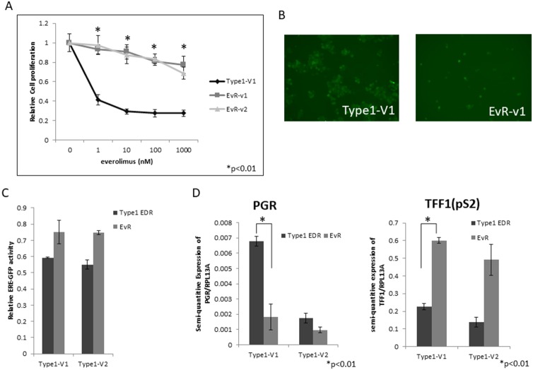
Figure 2. EvR cell establishment and characterization.
Effect of everolimus on Type 1 EDR cells (using V1) and everolimus-resistant variants established from Type 1 EDR-V1 (v1, v2) cells. (A) Everolimus-resistant cell (EvR) variants were cloned as follows: after long-term everolimus exposure and culture in a single dish, surviving cells were isolated and harvested. Highly proliferated variants were selected (variants 1 and 2). Everolimus suppressed cell proliferation in parental EDR cells, but not EvR cells. All data are shown as means ± SD of three independent experiments. *P<0.01 between parental Type 1 EDR and Type 1 EvR v1, v2 cells. GFP positivity was maintained after resistance to everolimus was acquired in Type 1 EDR cells (EvR-v1). (B) Comparison of luciferase assay in EDR and EvR cells. (C) Both Type 1 EDR (V1, V2) and EvR (generated individually from Type 1 EDR-V1 and V2) cells showed high estrogen response element-green fluorescent protein activity. PGR and TFF1 (pS2) expression in Type 1 EDR (V1, V2) and EvR cells. (D) Data are shown as means ± SD of three independent experiments. *P<0.01 between parental Type 1 EDR and Type 1 EvR v1 cells.

