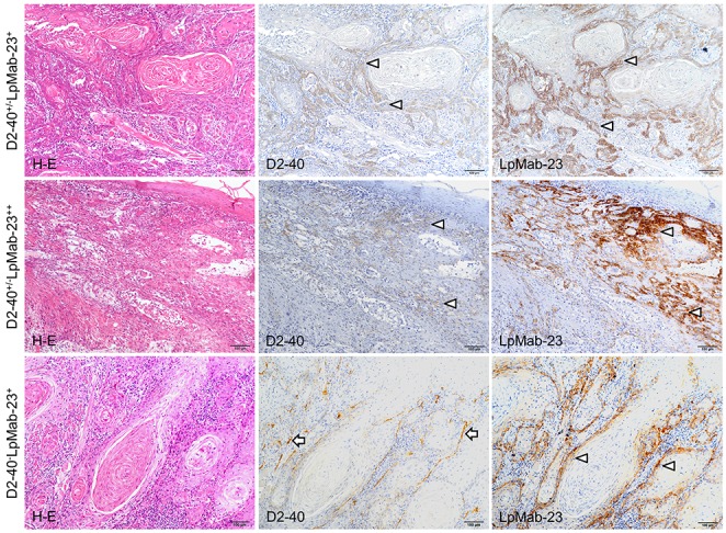
Figure 2. D2-40 dull/negative and LpMab-23 positive early stage tongue squamous cell carcinoma.

The sections are visualized by DAB staining for reaction products with D2-40 to normal PDPN and LpMab-23 for the cancer-specific PDPN with Hematoxylin staining of nuclei (blue). The top row shows panels of a D2-40 dull (middle) and LpMab-23-positive (right) tongue carcinoma (77 year old female, Grade 2, YK-4C, no recurrence, with metastasis). The cells of the cancer at the margins are weakly reacted with D2-40 (arrows) while in the adjacent section, the reaction with LpMab-23 (arrows) is stronger than that with D2-40. Bars: 100 μm. The middle row shows panels of a D2-40 dull and LpMab-23-positive tongue carcinoma (79 year old female, Grade 1, YK-4C, no recurrence, with metastasis). The invasive cancer cells showed few reactions with D2-40 (arrows) while in the adjacent section the cells showed strong reaction with LpMab-23 (arrows). Bars: 100 μm. The bottom row shows panels of a D2-40 negative and LpMab-23 positive-tongue carcinoma (77 year old female, Grade 2, YK-4C, no recurrence, with lymph node metastasis). The invasive cancer cells show no reactions with D2-40 while there is lymphangiogenesis with much D2-40-positive lymphatic vasculature found among cancer cells (arrowheads). In the adjacent section the cancer cells show a reaction with LpMab-23 (arrows). Bars: 100 μm.
