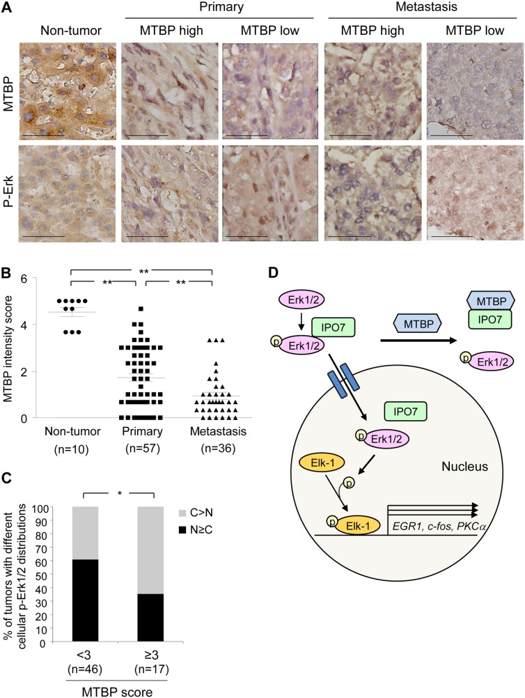
Figure 7. High MTBP levels are correlated with cytoplasmic p-Erk in human HCC tissues.
(A) Representative images of IHC for MTBP and p-Erk using human non-tumor liver tissues (n=10), primary HCC (n= 57), and metastatic HCC (n= 36) tissues. MTBP high: score≥3, MTBP low: score<3. Scale bar, 25 μm. (B) Summary of IHC for MTBP. Graph showing MTBP expression (immunoreactive) scores in non-tumor liver, primary HCC, and metastatic HCC tissues. Mann-Whitney test: **, P < 0.01. (C) Summary of association between MTBP levels and subcellular localization of p-Erk in human HCC tissues (combining primary and metastatic tissues). Graph showing % of tumors with different cellular distributions of p-Erk by expression scores of MTBP (<3 vs ≥3). Exact logistic regression controlling for tumor location: *, P < 0.05. (D) Proposed model for MTBP-mediated inhibition of the Erk1/2-Elk-1 signaling pathway. MTBP inhibits binding of p-Erk with IPO7, which reduces nuclear import of p-Erk, leading to decrease in Elk-1 phosphorylation and Elk-1 downstream target gene expression (EGR1, c-fos, PKCα).

