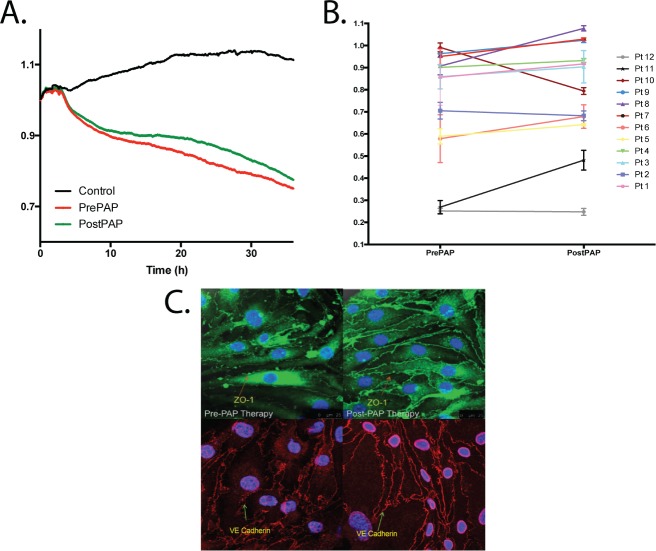
Figure 1. Exosome-mediated in vitro effects on endothelial cell monolayer resistance and membrane tight junction proteins.
(A) Ensemble-averaged curves of ECIS measured endothelial cell barrier resistance changes over time after administration of exosomes from adult patients with severe OSA/OHS before (Pre-PAP; red line) and after (Post-PAP; green line) positive airway pressure therapy compared to endothelial cells incubated with plasma free media and empty exosomes (control; black line). (The y axis refers to a ratio of ECIS values to baseline ECIS values at start of experiment). (B) Evaluation of ECIS measured endothelial cell barrier resistance changes of all 12 individual patients (pt) comparing the effects on endothelial cells 24 hours after exosome administration. ECIS changes were evaluated before (Pre-PAP) and after 6 weeks positive airway pressure therapy (Post-PAP). The overall cumulative improvement in endothelial cell monolayer resistance for all 12 subjects was +10.8% (P = .035). (C) Effect of plasma exosomes on tight junction and membrane structure in naive endothelial cells. Representative images of at least six separate experiments illustrate exosome-induced changes in expression of VE-cadherin and zonula occludens (ZO)-1 expression patterns. The scale bars for all the representative images are 25 μm. Left figures represent before and right figures represent after PAP therapy. ECIS = electric cell-substrate impedance sensing, OHS = obesity hypoventilation syndrome, OSA = obstructive sleep apnea, PAP = positive airway pressure.

