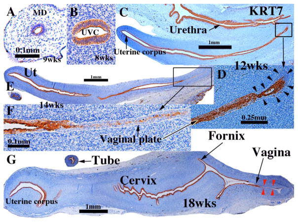
Fig. 2.
Keratin 7 immunohistochemistry of human fetal female reproductive tract development. (A & B) Müllerian duct and uterovaginal canal in 8 to 9-week human fetuses as indicated. (C & D) Sagittal sections of the human fetal female reproductive tract at 12 weeks of gestation. Note absence of staining of basal epithelial cells (arrowheads in D) at the caudal end of the vaginal plate. (E & F) Sagittal sections of the human fetal female reproductive tract at 14 weeks of gestation. Note continuous KRT7 staining from the uterine corpus to the solid vaginal plate, which shows reduced staining (F). (G) Sagittal section of the human fetal female reproductive tract at 18 weeks of gestation. KRT7 is expressed strongly in epithelium throughout the female reproductive tract except for the caudal portion of the vagina (red arrowheads).

