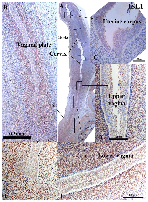
Fig. 8.
ISL1 immunohistochemistry of a 16-week human fetal female reproductive tract in sagittal sections. ISL1 is expressed in mesenchymal cells associated with the vaginal plate (A, B, E), the vagina (D & F), but not in the mesenchymal cells associated with the cervix (A), uterine corpus (A & C) and the uterine tube (not illustrated).

