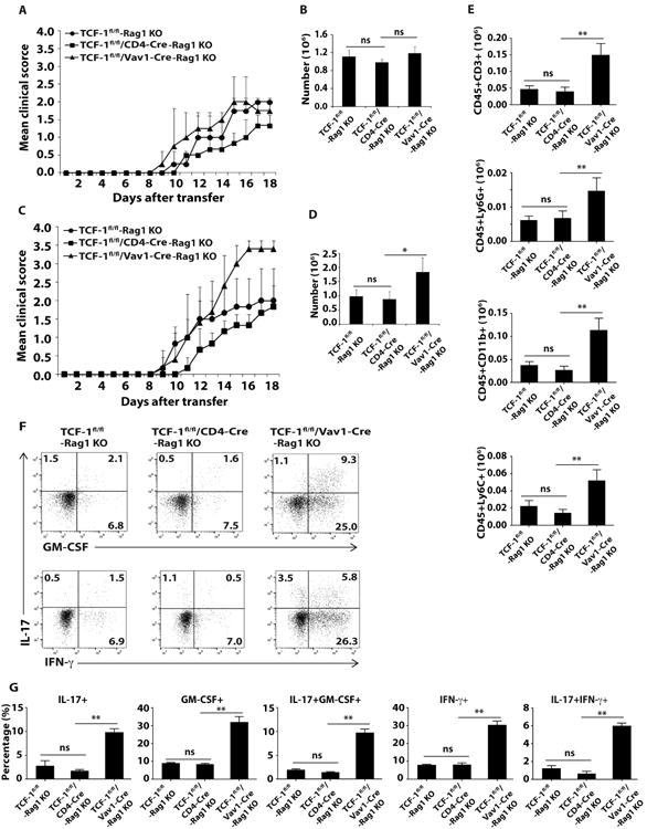
Figure 2.

Deletion of TCF-1 by Vav1-Cre potentiates Th17 but not Th1-mediated EAE. A, C, Mean clinical scores of EAE in Rag1-/- mice different days after adoptively transferred with indicated genotypes of MOG33–35-expanded Th1 (A) or Th17 (C) cells. B, D, Number of lymphocytes infiltrated into the CNS of Rag1-/- mice adoptively transferred with indicated Th1 (B) or Th17 (D) cells at the peak of disease as shown in A or C. E, Quantification of cells expressing surface markers characteristic of various types of lymphocytes as determined by flow cytometric analysis at the peak of disease from Rag1-/- mice receiving Th17 cells. F, Flow cytometric analysis of intracellular IL-17, GM-CSF and IFN-γ in CD4+ T cells that infiltrated CNS of Rag1-/- mice receiving Th17 cells at the peak of disease. G, Percentage of cells positive for indicated cytokines among CD4+ T cells infiltrated into CNS of Rag1-/- mice. Data are pooled from three experiments. ns, no significant difference; * < 0.05 and **<0.01.
