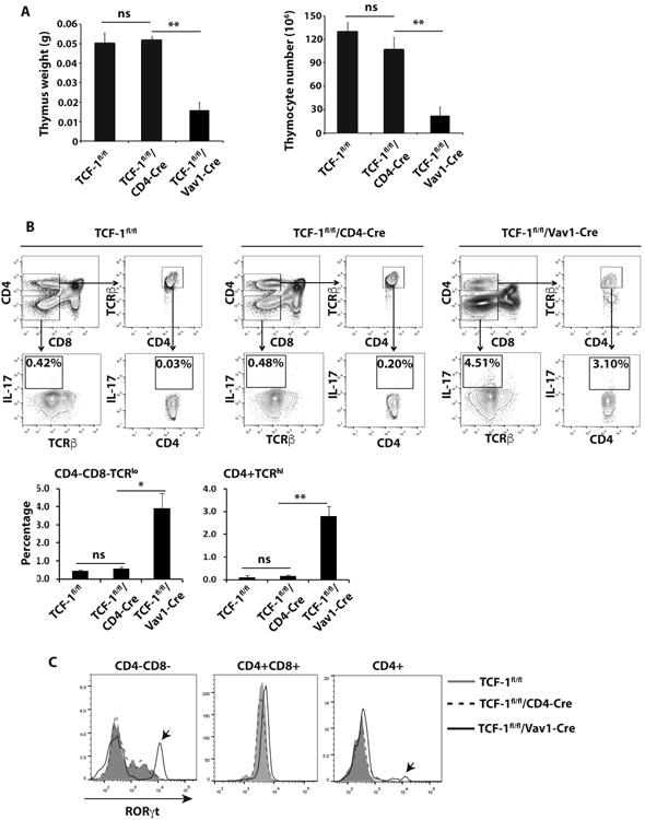
Figure 3.

Vav1-Cre induced deletion of TCF-1 leads to abnormal IL-17 expression in thymus. A, Weight (left panel) and number of total thymocytes (right panel) of the thymus (n=3) from indicated genotypes of mice. B, Thymocytes of different genotypes of mice were either gated on CD4-CD8- or CD4+ and TCRβhi (top panels). Percentage of IL-17+ cells in CD4-CD8- TCRβlo and CD4+ TCRβhi population of indicated genotype of mice was determined (middle panels). Bottom panels are the quantification of percentage of IL-17+ cells shown on middle panels from three mice of each genotype. C, Expression of RORγt in indicated population of cells from indicated genotypes of mice, detected by intracellular staining. Arrow indicates the very high levels of RORγt population in CD4-CD8- and CD4+ thymocytes of TCF-1f/fl /Vav1-Cre mice.
