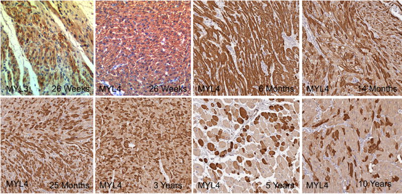
Fig. 4.

Age related changes in mosaicism. MYL3 was mosaic at 26 weeks in the atria, while MYL4 remained consistently expressed in the ventricle through 6 months and then decreased the percent of MYL4+ cells to 10 years of age (Original Magnification 20× for all images).
