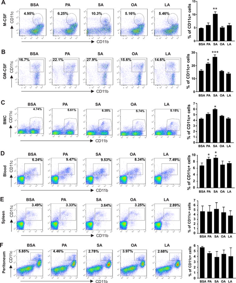
Figure 2. SA enhances CD11c+ macrophage differentiation in vitro.

(A-B) Bone marrow cells collected from 6-8 weeks old C57BL/6 mice were differentiated into macrophages under stimulation of M-CSF (20ng/ml) (A) or GM-CSF (20ng/ml) (B) in the presence of individual FAs, including PA (100μM), SA (100μM), OA (100μM) and LA (100μM), and BSA control for 3 days. Flow cytometric staining for analysis of CD11c expression in CD11b+ cells. Average percentage of CD11c+ macrophages is shown in the right panel. (C-F) Cells from bone marrow (BMC) (C), peripheral blood (D), spleen (E) and peritoneum (F) collected from 6-8 weeks old C57BL/6 mice were directly cultured in vitro with PA(100μM), SA(100μM), OA(100μM), LA(100μM) or BSA controls for 36h and flow cytometric analysis of CD11c expression on CD11b+ populations. Average percentage of CD11c+ cells is shown in the right panel. Data are mean value ± SEM of three mice (* p<0.05, ** p<0.01, *** p<0.001 compared to BSA controls), and are representative of three independent experiments.
