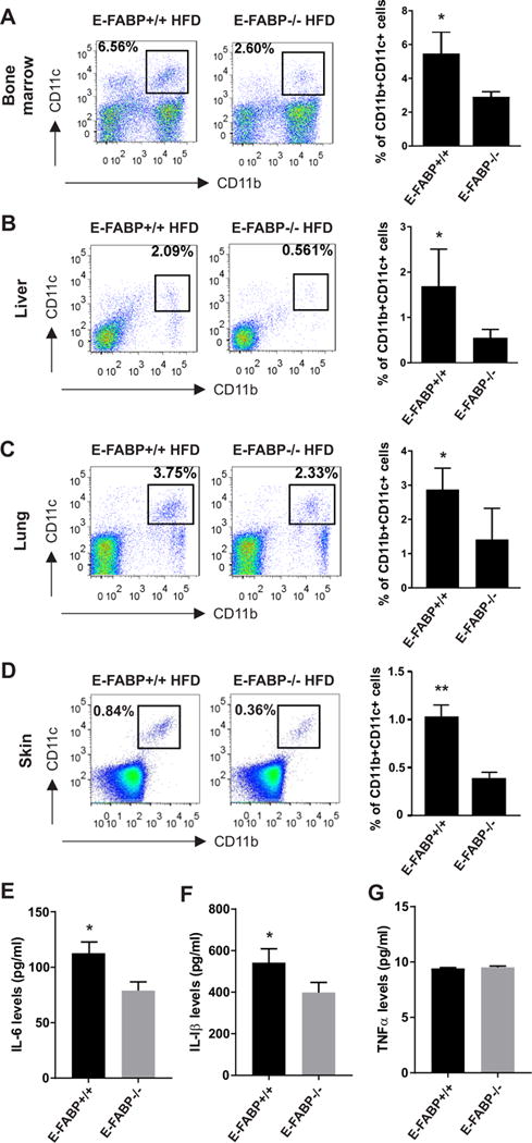
Figure 6. E-FABP deficiency reduces CD11c+ macrophages in obese mice.

WT and E-FABP−/− mice were fed on HFD (60% fat) for 20 weeks. Different tissues or organs were collected, respectively from the obese WT and E-FABP−/− mice (n=5) for analysis of the presence of CD11b+CD11c+ macrophages. (A-D) Flow cytometric surface staining for analysis of CD11b+CD11c+ macrophages in bone marrow (A), liver (B), lung (C) and skin (D). Average percentage of CD11c+ cells is shown in the right panel. (E-G) Measurement of cytokine levels of IL-6 (E), IL-1β (F) and TNFα (G) in the serum collected from obese WT and E-FABP−/− mice by ELISA. Data shown as mean ± SEM (*p<0.05, **p<0.01), and are representative of at least two independent experiments. Also see Figure S2.
