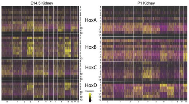
Fig. 1. Hox gene expression in the developing kidney shows little evidence for paralog Hox codes.
The expression patterns for all Hox genes in the compartments of the early developing E14.5 and early postnatal (P1) kidney. The extreme terminal Hox genes from paralog groups 1 and 13 showed very low expression. For the HoxA, HoxB and HoxC clusters all of the genes of a given cluster showed very similar expression patterns, although with varying expression levels. For example, almost all of the HoxB genes, including paralog groups 1,2,3,4,5,6,7,8,9,13, showed stronger expression in cell types 1,3,4,8,9 and 11 and weaker expression in the other compartments. The HoxD cluster was an exception, with paralog Hox gene specific expression patterns in cell types 3,4,5,9 and 10. Cell type identities for E14.5 are 0, Stroma: 1, Differentiating Nephrons: 2, Medullary Stroma: 3, Loop of Henle: 4, Cap Mesenchyme: 5, Stromal Progenitors: 6, Endothelium: 7, Podocytes: 8, Cap Mesenchyme: 9, Collecting Duct: 10, Cortical Stroma: 11, Cap Mesenchyme: 12, Immune Cells. P1 compartments are as follows: 0, Nephron Progenitors: 1, Proximal Tubule: 2, Early Proximal Tubule: 3, Loop of Henle: 4, Cap Mesenchyme: 5, Stroma: 6, Collecting Duct: 7, Distal Tubule: 8, Endothelium: 9, Podocytes: 10, Collecting Duct Tip Cells. Only Hox genes with some measured expression in the developing kidney are included. Genes with no detected expression included at E14.5 Hoxc12 and Hoxd4, and at P1 Hoxb13, Hoxc12,13, and Hoxd4,13.

