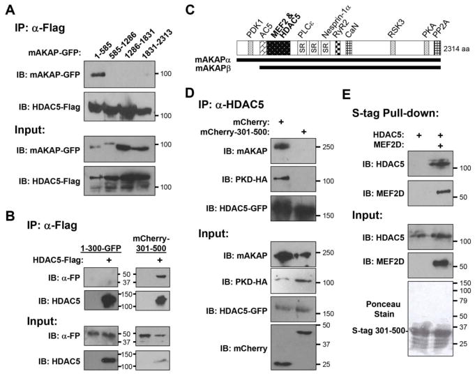
Figure 2. HDAC5 is associated with a discrete mAKAP domain.
A, B. Extracts prepared from HEK293 cells co-transfected with expression plasmids for Flag-tagged HDAC5 and GFP- or mCherry tagged mAKAP fragments were used in co-immunoprecipitation assays with Flag tag antibodies. Expression of the recombinant proteins was detected using tag-specific antibodies. FP - fluorescent protein, i.e. GFP or mCherry. C. mAKAP domain structure. mAKAPβ is identical to mAKAPα 245–2314 [54]. SR - spectrin repeat domain. Binding sites are indicated for those mAKAP-binding partners whose sites have been finely mapped: PDK1, protein kinase D 1 [54]; AC5, adenylyl cyclase 5 [40]; PLCε, phospholipase C ε [22]; nesprin-1α [23]; RyR2, ryanodine receptor [55]; CaN, calcineurin [56]; RSK3, p90 ribosomal S6 kinase 3 [57]; PKA [18]; PP2A, protein phosphatase 2A [58]. D. Cardiac myocytes were infected with adenovirus expressing HA-tagged PKD, GFP-tagged HDAC5, mCherry or mCherry-tagged mAKAP 301–500. Protein complexes immunoprecipitated with HDAC5 antibodies. E. Bacterially-expressed His-tagged HDAC5 and MEF2D and S-tagged mAKAP 301–500 were used in pull-down assays with S-tag resin. S-tagged mAKAP 301–500 was detected by total protein stain of the pull-downs (Ponceau stain). n = 3 for each panel.

