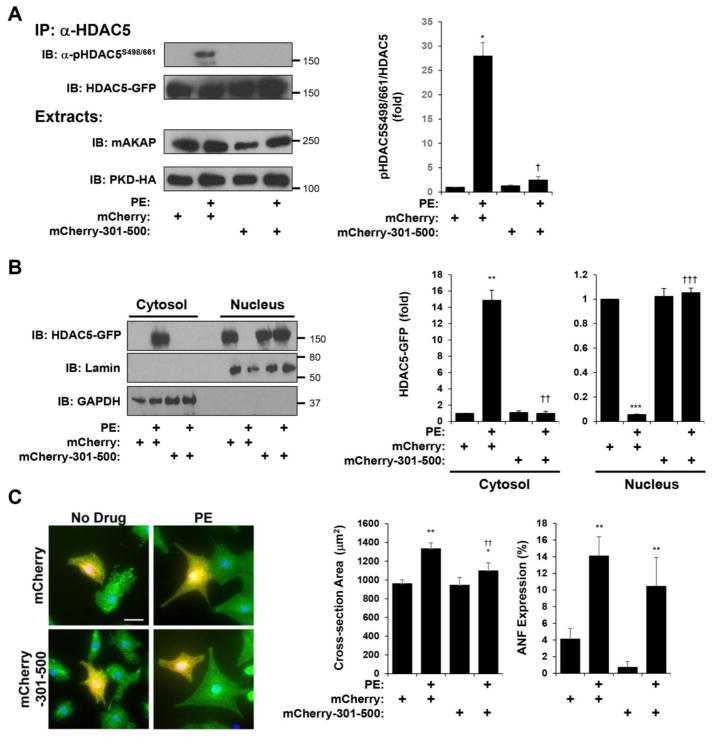
Figure 3. HDAC5 association with mAKAPβ signalosomes is required for α-adrenergic-induced HDAC5 nuclear export and neonatal myocyte hypertrophy.
A. Cardiac myocytes were infected with adenovirus expressing HDAC5-GFP, PKD-HA and either mCherry-mAKAP 301–500 or mCherry control before stimulation with PE (50 μM) for 60 min. HDAC5-GFP was immunoprecipitated with a HDAC5 antibody and detected using a phospho-specific antibody for Ser-498/661. B. Cytosolic and nuclear extracts were prepared from myocytes treated as in A and analyzed by western blotting with HDAC5, lamin (nuclear marker), or GAPDH (cytosolic marker) antibodies. For A and B, n = 3. C. Cardiac myocytes were transfected with ptTA and pTRE expression plasmids for either mCherry-mAKAP 301–500 or mCherry control before stimulation with PE (10 μM) for 2 days and staining with α-actinin antibody (green) and Hoechst nuclear stain (blue). Parallel cultures were stained for ANF expression (not shown). n = 5 separate biological replicates. *p-value vs. no drug controls; † vs. mCherry + PE. For ANF expression, 2-way matched ANOVA was significant for anchoring disruptor expression (p = 0.03 for samples with mCherry-301-500 vs. samples with mCherry control), albeit pairwise post-hoc testing comparing PE-treated control and 301–500 peptide samples did not reach statistical significance.

