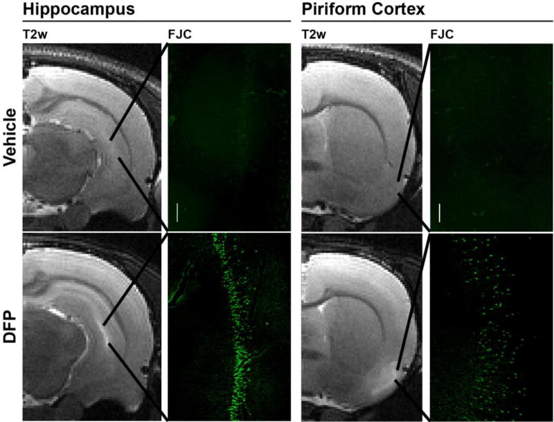
Figure 3. Lesions detected by T2w MRI at 72 h post DFP intoxication correspond to regions of ongoing neurodegeneration as assessed by FluoroJade C (FJC) staining.
The vehicle control animal shows no T2w hyperintensity or FJC positive neurons in the hippocampus or piriform cortex. In contrast, DFP intoxication produced hyperintensity in the hippocampus and piriform cortex that shows spatial registry to bands of FJC positive cells. Magnification: 100×.

