
Figure 4.
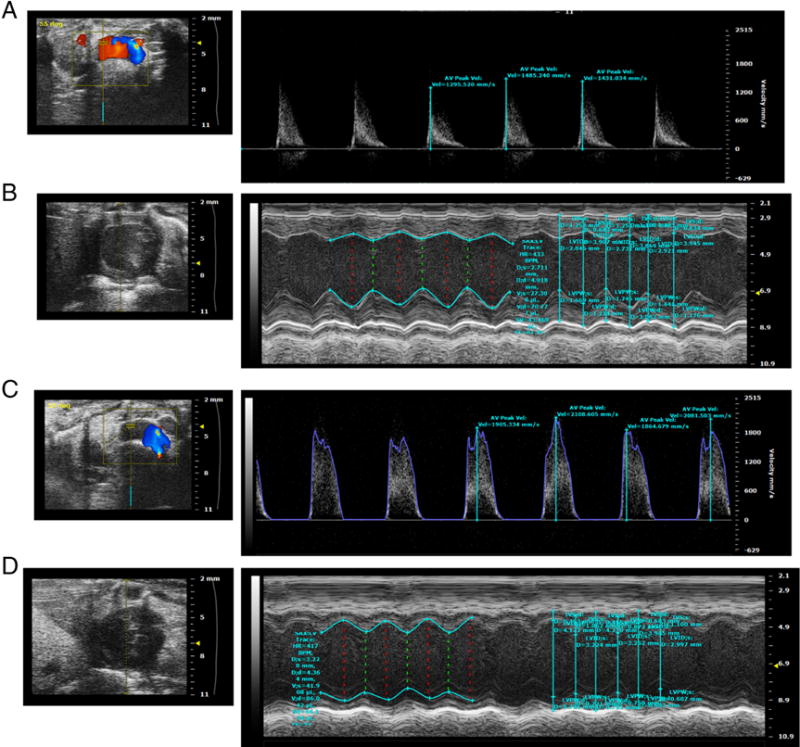
Aortic Valve Echo Doppler (A), and B mode and M mode left ventricle views (B) of a representative NC fed LDLr−/−ApoB100/100 mouse. Aortic Valve Echo Doppler (C), and B mode and M mode left ventricle views (D) of a representative DB fed LDLr−/−ApoB100/100 mouse.