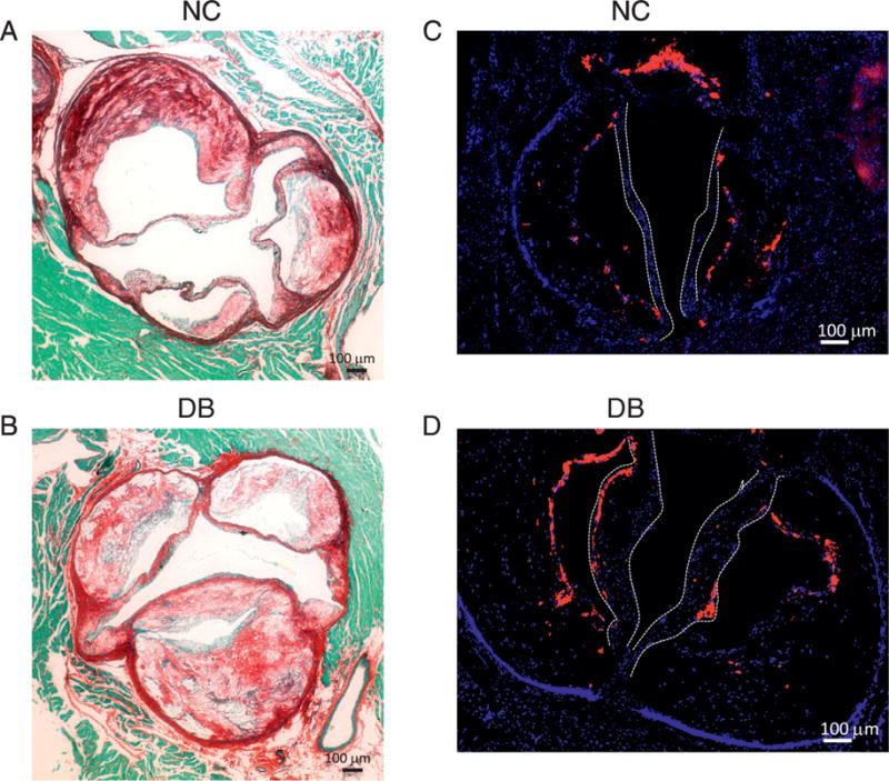
Figure 7.

(A and B) histological staining for Picrosirus Red (A and B) showing extensive collagen fibers throughout the aortic sinus and in the thickened valve leaflets in DB fed mice (B). (C and D) immunofluorescence staining for the Mac2 antigen labeling macrophages showing extensive macrophage accumulation in the leaflets of DB fed mice. Leaflets shown by white dotted lines.
