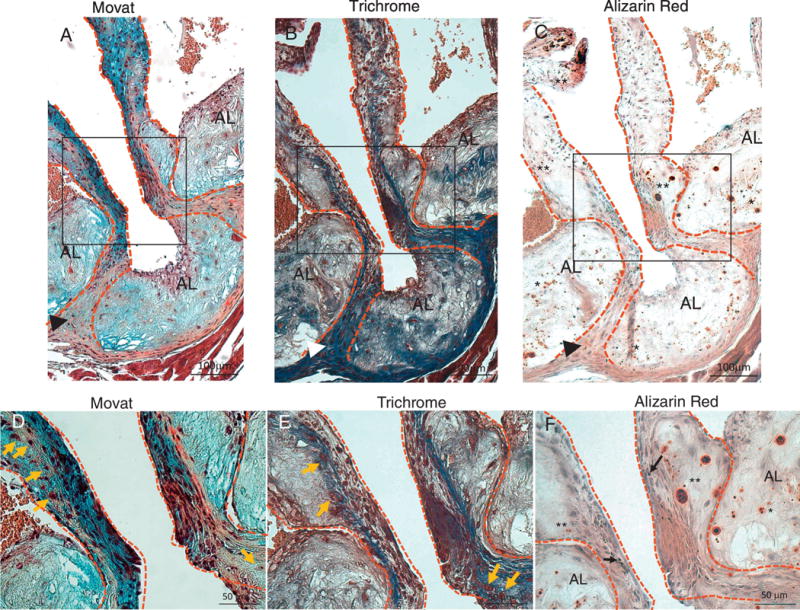
Figure 8.

Movat (A and D), Trichrome (B and E), and Alizarin Red (C and F) staining of aortic sinus adjacent sections from DB fed mice. D, E and F are higher magnification of insets in A, B and C respectively. Dotted lines contour the valve leaflets and hinge areas. (A–C) Chondrocyte-like cells in hinge area co-localize with dense collagen and calcium deposits (arrow-heads). Extensive calcification in the leaflets (**, C and F) is associated with collagen and proteoglycan rich areas. Extensive calcification is also present in the atherosclerotic lesion (AL) (*, C and F) associated with collagen and proteoglycan rich areas. Chondrocyte-like cells are also present in the leaflets and hinge areas (orange arrows, D and E). Melanocytes (black arrows, F) are present in the leaflets. AL=atherosclerotic lesion areas, **=leaflet calcification, *=atherosclerotic lesion calcification, arrow-head=calcification and chondrocyte cells in the hinge areas, orange arrows=chondrocyte-like cells.
