Figure 1.
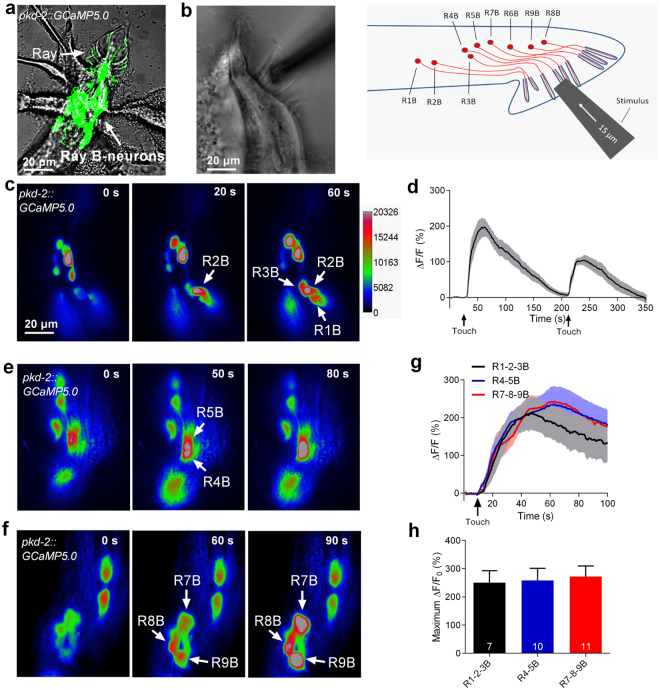
Touch-induced calcium responses in RnB neurons. (a) Micrograph of the male tail showing the expression of pkd-2::GCaMP5.0 in the rays. All RnB neurons, except R6B, express GCaMP5.0 by the control of the pkd-2 promoter. (b) A schematic illustrating of delivering mechanical stimulation toward the RnB cilia (at the position of rays 4–6 is shown). Worms expressing GCaMP5.0 in RnB neurons were immobilized with glue and immersed in a bath solution. (c) Representative time-lapse rainbow images of GCaMP5.0 based calcium responses from R1B- R3B neurons induced by mechanical stimulation of 15 μm displacement at the position of rays 1–3. (d) Calcium responses of the R1B- R3B neurons induced by two successive mechanical stimuli of 15 μm displacement with 180 s interval at the position of rays 1–3. Solid lines show the average fluorescence changes and the shading indicates SEM. n = 10. (e) Representative time-lapse rainbow images of GCaMP5.0 based calcium responses from R4B and R5B induced by mechanical stimulation of 15 μm displacement at the position of rays 4, 5. (f) Representative time-lapse rainbow images of GCaMP5.0 based calcium responses GCaMP5.0 based calcium responses from R7B- R9B neurons induced by mechanical stimulation of 15 μm displacement at the position of rays 7–9. (g,h) Calcium responses (g) and maximum ΔF/F0 changes (h) of the RnB neurons in response to mechanical stimulation. Solid lines show the average fluorescence changes and the shading indicates SEM. n ≥ 7. All error bars represent SEM.

