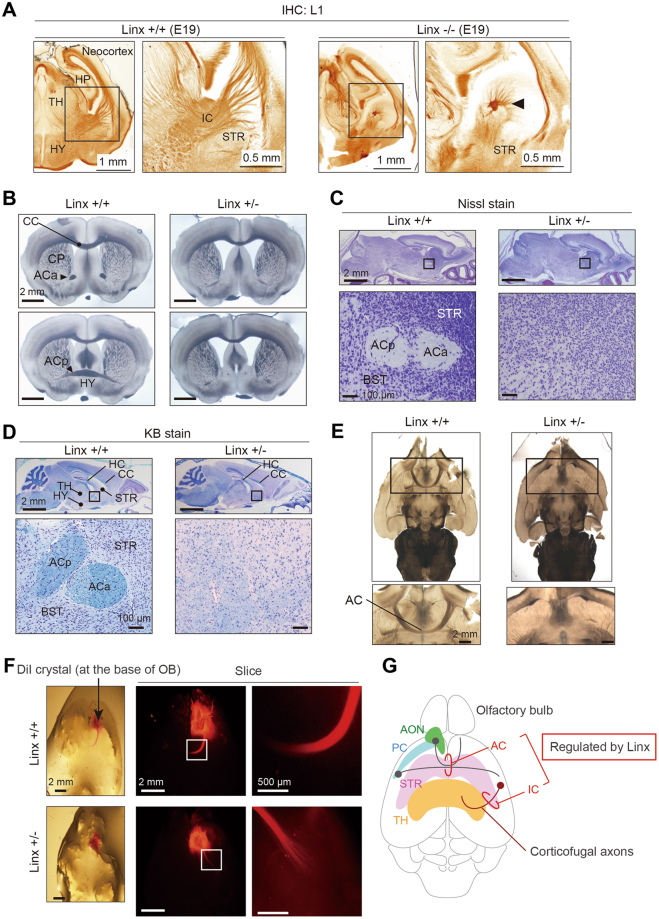
Figure 3.
Defective AC development in Linx+/− mice. (A) A defect in IC development found in Linx−/− mice. The coronal sections of brains from Linx+/+ (left) and Linx−/− (right) E19 embryonic mice were stained for L1, a neuronal adhesion molecule, to visualize the IC. An arrowhead denotes the remnants of the descending component of the IC. Boxed areas were magnified in the adjacent panels. (B–D) Transmitted light (B), Nissl-stained (C), and KB-stained (D) images of coronal sections from P30 mouse brains showing the defects in the AC of Linx+/− mice. ACp, posterior branch of the AC; ACa, anterior branch of the AC; TH, thalamus; HC, hippocampal commissure; BST, bed nuclei of the stria terminalis. (E) Horizontal sections from the brains of wild-type (left) and Linx+/− (right) mice showing defective AC development in Linx+/− mice. Boxed areas are magnified in the lower panels. (F) The axon tract from the AON was labeled with DiI crystals placed around the AON regions prepared from wild-type and Linx+/− mice. The axon tract that constitutes the anterior branch of the AC was short and disrupted in Linx+/− mice compared with wild-type mice. Boxed areas are magnified in the adjacent panels. (G) A schematic illustration representing the neural projections regulated by Linx; this is based on data from both previous12 and present studies.

