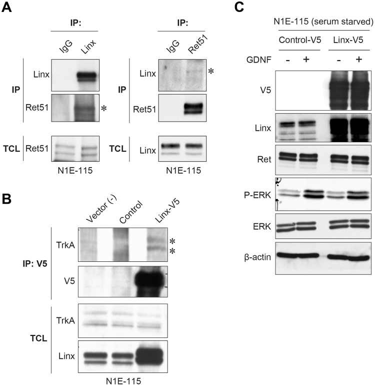
Figure 5.
Interaction of Linx with RTKs and its effect on ERK signaling. (A) Linx interaction with Ret. Lysates from N1E-115 cells were immunoprecipitated (IP) using Linx (left) and Ret51 (right; an isoform of Ret) antibodies, followed by Western blot analysis with the indicated antibodies. Asterisks indicate co-immunoprecipitated Ret51 and Linx. TCL, total cell lysates. Full blot images are shown in Supplementary Figure S3. (B) Linx interaction with TrkA. Lysates from N1E-115 cells transfected with either control or Linx-V5 vector were immunoprecipitated with V5 antibody, followed by Western blot analysis. Asterisks indicate co-immunoprecipitated TrkA. Full blot images are shown in Supplementary Figure S3. (C) N1E-115 cells transfected with either control or Linx-V5 vector were starved and stimulated with GDNF, followed by Western blot analysis using the indicated antibodies. Full blot images are shown in Supplementary Figure S3.

