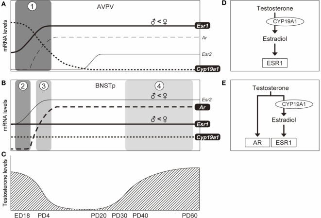
Figure 6.
Schematic illustration of the temporal expression patterns of genes in the AVPV and BNSTp and possible testicular testosterone actions on the formation of the AVPV and BNSTp in male mice. In the AVPV [panel (A)], based upon our previous (23) and current studies, Esr1 mRNA expression (solid line) increases from embryonic day 18 (ED18) to PD4 and continues in the pubertal period. Cyp19a1 mRNA level (dotted line) decreases from ED18 to PD4 and is not expressed by PD20. Esr1 and Cyp19a1 are essential to the formation of the male AVPV, but Esr2 and Ar are not necessary (23). Testicular testosterone production reaches a peak level at ED18 and decreases after birth (50), and again is increased from PD35 (44) [panel (C)]. The temporal patterns of Esr1, Cyp19a1, and testicular testosterone may indicate that the male AVPV is organized by estradiol (formed by the aromatization of testosterone) signaling through ESR1 during the perinatal period [boxed phase 1 in panel (A) and panel (D)]. In the BNSTp [panel (B)], based upon our previous (23) and current studies, Esr1 mRNA (solid line) is expressed during the perinatal period, and is also expressed during the pubertal period. Cyp19a1 mRNA level (dotted line) is stably expressed in the perinatal and pubertal periods. Ar mRNA (broken line) is not expressed in the late fetal period (ED18), but expressed in the postnatal (PD4) and pubertal periods. Esr1, Cyp19a1, and AR are requisite for the sexual differentiation of the male BNSTp (23, 26). The patterns of Esr1, Cyp19a1, Ar, and testicular testosterone levels may indicate that the male BNSTp is formed by the action of estradiol (formed by the aromatization of testicular testosterone) via ESR1 in the fetal period [boxed phase 2 in panel (B) and panel (D)], and both the action of estradiol via ESR1 and testicular testosterone via AR in the postnatal [boxed phase 3 in panel (B) and panel (E)] and pubertal [boxed phase 4 in panel (B) and panel (E)] periods.

