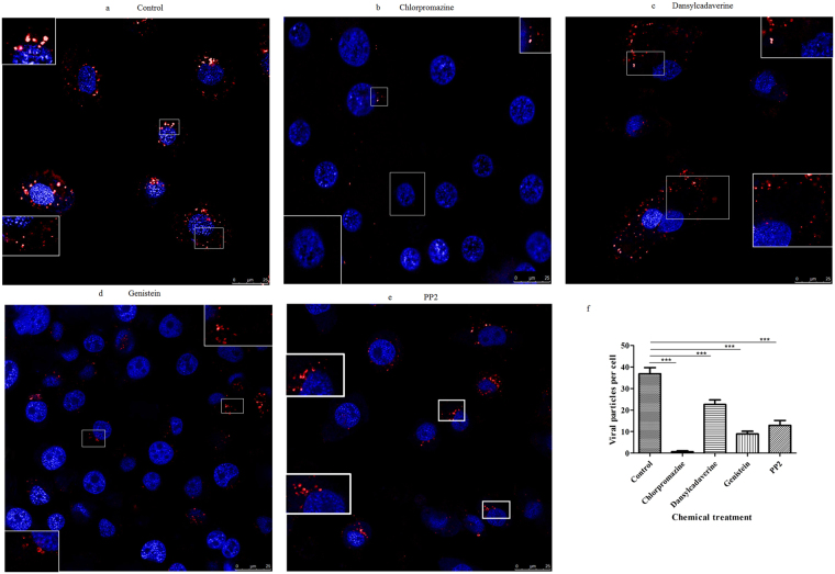
Figure 3.
Confocal microscopy observations of BmCPV internalization and different distribution of BmCPV particls with different endocytic inhibitors. BmN cells were treated without inhibitors as a control (a); the cells were treated with chlorpromazine (2 mM) (b), dansylcadaverine (2 mM) (c), genistein (50 μg/mL) (d), or PP2 (0.16 μM) (e), for 30 min or 1 h, followed by inoculation with A546-labeled BmCPV virions. Thirty minutes after inoculation, images were captured by a confocal microscopy. The nucleus was stained withDAPI. (f) Quantification of internalized BmCPV particles in single planes of view (n = 21 cells). Error bars indicate standard deviations. ***P < 0.001.

