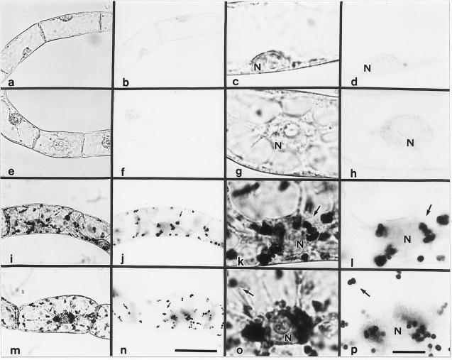
Figure 3.
Photomicrographs of amyloplasts in BY-2 cultured cells stained with iodine. Stationary-phase (a–d), 2-d-old cells cultured in D medium (e–h), F medium (i–l), or B medium (m–p) are shown. a, c, e, g, i, k, m, and o, Cells observed with the condenser diaphragm closed; b, d, f, h, j, l, and p, cells observed with the condenser diaphragm open. a and b, c and d, e and f, g and h, i and j, k and l, m and n, and o and p show the same fields. a, b, e, f, i, j, m, and n and c, d, g, h, k, l, o, and p are at the same magnification. Arrows indicate starch granules. N, Cell nucleus. The bars in n and p represent 50 and 10 μm, respectively.

