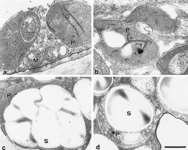
Figure 4.
Electron micrographs of leucoplast-like plastids in stationary-phase cells (a), proplastids observed in cells cultured in D medium for 48 h (b), and differentiated amyloplasts in 2-d-old cells cultured in F medium (c) and B medium (d), which are filled with starch grains. M, Mitochondrion; P, plastid; S, starch granule. Scale bar represents 500 nm.

