Figure 5.
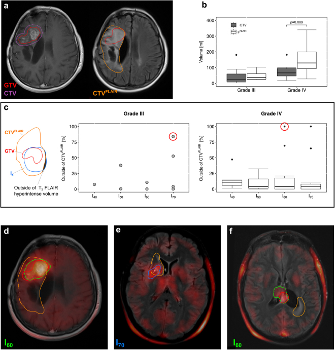
Coverage of 18F-FET PET isocontour volumes by T2 FLAIR hyperintense regions. Novel guidelines suggest inclusion of regions that are hyperintense on T2 FLAIR MRI (orange) into CTVFLAIR (a). In grade IV glioma, this significantly increases the volume of CTV (b). The fraction of the 18F-FET PET based isocontour volumes that lies outside of the T2 FLAIR hyperintense volume, was calculated (c). With two exceptions for isocontours of 70% in grade III tumors and three exceptions for isocontours of either 60% or 70% in grade IV tumors, more than 80% of the isocontours were covered by the T2 FLAIR hyperintense volume (c). Representative patient with grade IV glioma where over 90% of the 18F-FET PET active area lies within CTVFLAIR (d). Exemplary cases of two exceptional patients circled in red in the boxplots (c) with large fractions of the isocontour volumes (70% and 60%) laying outside the T2 FLAIR hyperintense volume are shown.

