Figure 2.
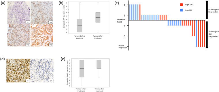
XPF and MUS81 protein levels in EAC tissue by immunohistochemistry. (a) Esophageal adenocarcinoma pre-treatment biopsies stained for XPF protein, photographed at x20 magnification; intensity scores (i) 0, (ii) 1, (iii) 2 and (iv) 3. (b) Significant increase in XPF protein levels when pre-treatment biopsies were compared with post-treatment resection samples following 2 cycles of oxaliplatin-fluorouracil chemotherapy (median pre-treatment 2, IQR 0–4; median post-treatment 4.5, IQR 3–6; P = 0.004, Wilcoxon signed rank test). (c) A waterfall plot of the association between pre-treatment tumour XPF level and pathological response following oxaliplatin-chemotherapy. Each line represents the degree of Mandard regression in an individual patient. Patients exhibiting Mandard grades 1–3 were classified as pathological responders; those with Mandard grades 4 and 5 were classified as non-responders10. Low XPF expression was associated with pathological response (odds ratio 3.85, P = 0.041). (d) MUS81 staining of esophageal adenocarcinoma pre-treatment biopsies, photographed at X20 magnification, showing examples of (i) high and (ii) low expression. (e) Trend towards increased MUS81 staining when pre-treatment biopsies were compared with post-treatment resection samples following 2 cycles of oxaliplatin-fluorouracil chemotherapy (median pre-treatment 6, IQR 3–9; median post-treatment 9, IQR 6–9; P = 0.051, Wilcoxon signed rank test).

