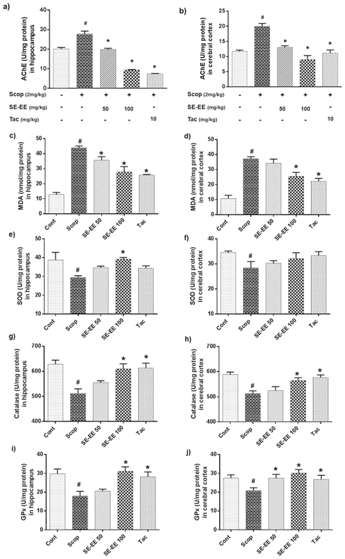
Figure 5.
Effects of SE-EE on scopolamine-inflicted cholinergic impairment, lipid peroxidation and oxidative stress in C57/BL6N mice: AChE activity – (a) AChE levels in hippocampus, (b) AChE levels in cerebral cortex. Tacrine (10 mg/kg) was used as positive control; Endogenous level of oxidative stress/antioxidant biomarkers – (c,e,g and i) represents the lipid peroxidation (MDA), Superoxide dismutase (SOD), Catalase (CAT) and Glutathione peroxidase (GPx) levels in the hippocampus respectively; (d,f,h and j) represents the MDA, SOD, CAT and GPx levels in the cerebral cortex of the treated animals, respectively. The data are expressed as mean ± SD (n = 5, pooled biological replicates). One-way ANOVA-Tukey’s multiple comparison test was performed. #p < 0.05 compared with the control group; *p < 0.05 other treated groups compared with the scopolamine group.

