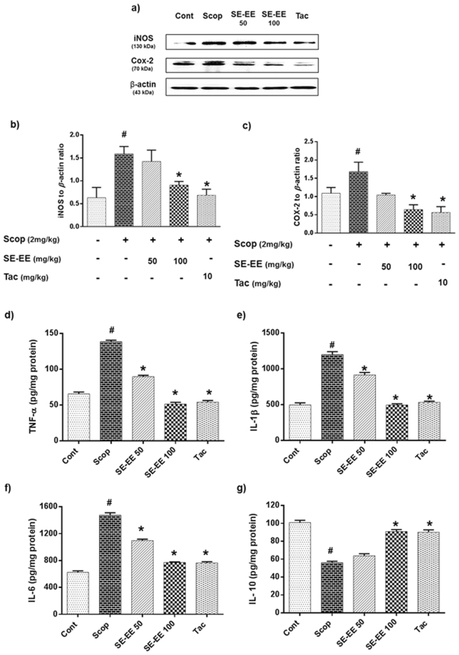
Figure 6.
Effects of SE-EE on scopolamine inflicted neuroinflammation in C57/BL6N mice: (a) iNOS and COX-2 protein expressions in the hippocampus (n = 3) was determined by western blotting analysis (respective markers were acquired by cropping from the same gel, full length blots are in Supplementary data- S.3.2) and (b,c) represents the quantification of inflammatory protein expression relative to β-actin, which was achieved using the ImageJ software; (d,e,f and g) represent the level of TNF-α, IL-1β, IL-6 and IL-10 proteins in the hippocampus (n = 5, pooled biological replicates), respectively, as acquired via ELISA. Data are expressed as mean ± SD. One-way ANOVA-Tukey’s multiple comparison test was performed. #p < 0.05 compared with the control group; *p < 0.05 other treated groups compared with the scopolamine group.

