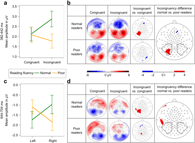
Figure 3.
EEG analyses. Mean amplitude values were calculated for electrodes of interest, marked with black polygons (n = 28). Error bars illustrate standard error of the mean. (a) Significant interaction of congruency and reading fluency for mean amplitude values in the time window 382–442 ms. (b) Potential field maps and statistical t-maps of initial ERP of audiovisual integration (382–442 ms). (c) Significant interaction of hemisphere and reading fluency for mean amplitude values in the time window 644–704 ms. (d) Potential field maps and statistical t-maps of late negativity ERP (644–704 ms).

