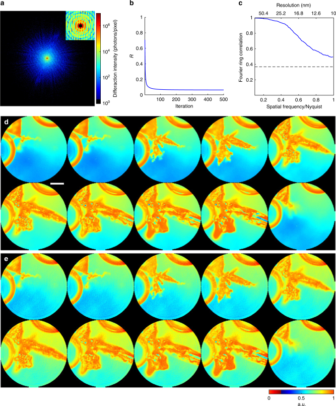
Fig. 2.
Numerical simulations of in situ CDI with coherent X-rays. a A representative diffraction pattern with Poisson noise and missing data, calculated from the Pb dendrite formation process in an electrochemical cell using 8 keV X-rays with a flux of 1011 photons μm−2 s. The insert indicates a 5 × 5 pixels missing data at the center. b R-factor used to monitor the iterative algorithm, showing the rapid convergence of the algorithm. c Average Fourier ring correlation between a time-evolving structure model and its corresponding reconstructions indicates a spatial resolution of 10 nm can be achieved, with a temporal resolution of 10 ms. d The time-evolving structure model of the dendrite formation process immersed in a 1-μm-thick water layer. Scale bar, 200 nm. e The corresponding reconstructions of the time-evolving complex exit waves (showing only the magnitude), which are in good agreement with the structure model

