Fig. 2.

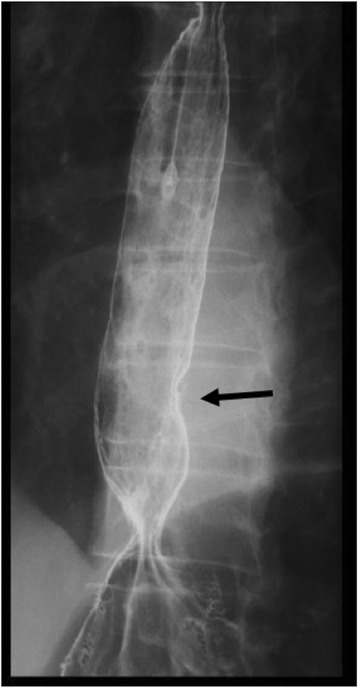
Esophagography. Esophagography showed a type 0-IIa + IIc elevated lesion (15 mm in size) on the left wall of the lower esophagus, and the tumor exhibited arcuate change suggesting submucosal invasion

Esophagography. Esophagography showed a type 0-IIa + IIc elevated lesion (15 mm in size) on the left wall of the lower esophagus, and the tumor exhibited arcuate change suggesting submucosal invasion