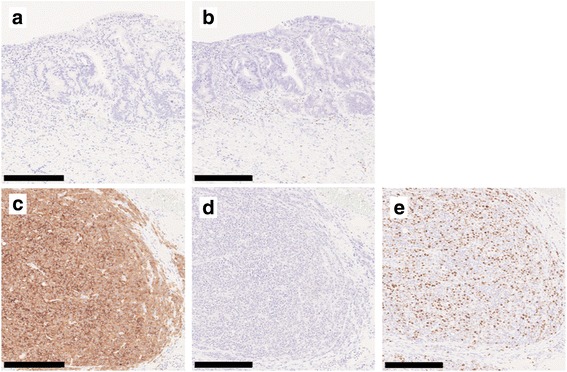
Fig. 5.

Immunohistochemical findings. Immunohistochemically, adenocarcinoma cells were negative for synaptophysin (a) and chromogranin A (b), while the round-shaped carcinoma cells were diffusely positive for synaptophysin (c), but negative for chromogranin A (d). The Ki67 labeling index was 50% (e). All scale bars—250 μm
