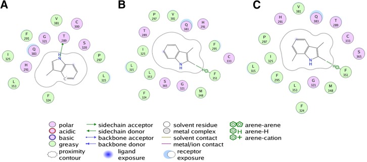
Fig. 9.
Schematic representation of AhR ligand binding domain docked with antagonists 3-Me-indole (A), 2,3-diMe-indole (B), and 2,3,7-triMe-indole (C). All antagonists bind to the same pocket as the agonists and share a conserved hydrogen bond interaction between the amine group and threonine 289 or arene interactions with aromatic residues in the pocket. All residues that lie within a 5 Å radius from the center of the binding pocket are listed and colored according to their amphiphilicity profile. The schematic legend details the nature of the interactions of antagonists with the residues in the binding pocket. The images were generated using the ligand interactions module in the MOE program.

